clitoris

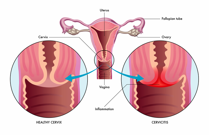
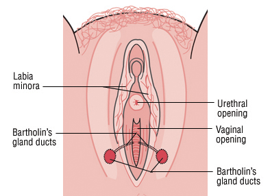
The clitoris is a female sex organ present in mammals, ostriches and a limited number of other animals. In humans, the visible portion – the glans – is at the front junction of the labia minora (inner lips), above the opening of the urethra. Unlike the penis, the male homologue (equivalent) to the clitoris, it usually does not contain the distal portion (or opening) of the urethra and is therefore not used for urination. The clitoris also usually lacks a reproductive function. While few animals urinate through the clitoris or use it reproductively, the spotted hyena, which has an especially large clitoris, urinates, mates, and gives birth via the organ. Some other mammals, such as lemurs and spider monkeys, also have a large clitoris.

Image result for Clitoris

For More Views: http://www.sciaeon.org/womens-health-and-complications/home        

Article page: http://www.sciaeon.org/womens-health-and-complications/articles-in-press

Contribute your manuscript: http://www.sciaeon.org/submit-paper

For more queries: whc@sciaeonopenaccess.com

Cystocele

Cystocele. A dropped or prolapsed bladder (cystocele) occurs when the bladder wall bulges into the vaginal space. It results when supporting muscles and tissues for the bladder give way. A cystocele, also called a prolapsed or dropped bladder, is the bulging or dropping of the bladder into the vagina. The bladder, located in the pelvis between the pelvic bones, is a hollow, muscular, balloon-shaped organ that expands as it fills with urine. During urination, also called voiding, the bladder empties through the urethra, located at the bottom of the bladder. The urethra is the tube that carries urine outside of the body. The vagina is the tube in a woman’s body that runs beside the urethra and connects the womb, or uterus, to the outside of the body.

Image result for cystocele

For More Views: http://www.sciaeon.org/womens-health-and-complications/home        

Article page: http://www.sciaeon.org/womens-health-and-complications/articles-in-press

Contribute your manuscript: http://www.sciaeon.org/submit-paper

For more queries: whc@sciaeonopenaccess.com

Cervicitis

Cervicitis is an inflammation and irritation of the cervix. Symptoms of cervicitis can be similar to vaginitis, with vaginal discharge, itching or pain with intercourse. Cervicitis can be caused by a sexually transmitted infection. Most common are chlamydia and gonorrhea. The cervix is the donut-shaped opening to the uterus. Cervicitis is an inflammation and irritation of the cervix. Symptoms of cervicitis can be similar to vaginitis, with vaginal discharge, itching or pain with intercourse. Cervicitis can be caused by a sexually transmitted infection. Most common are chlamydia and gonorrhea. Trichomoniasis and genital herpes can also cause cervicitis. In some cases, cervicitis is not caused by infection. It may be due to trauma, frequent douching or exposure to chemical irritants.

Image result for cervicitis

For More Views: http://www.sciaeon.org/womens-health-and-complications/home        

Article page: http://www.sciaeon.org/womens-health-and-complications/articles-in-press

Contribute your manuscript: http://www.sciaeon.org/submit-paper

For more queries: whc@sciaeonopenaccess.com

Catheter

A urinary catheter tube drains urine from your bladder. You may need a catheter because you have urinary incontinence (leakage), urinary retention (not being able to urinate), prostate problems, or surgery that made it necessary. Clean intermittent catheterization can be done using clean techniques. In medicine, a catheter is a thin tube made from medical grade materials serving a broad range of functions. Catheters are medical devices that can be inserted in the body to treat diseases or perform a surgical procedure. Insertion and removal of poorly lubricated catheters causes friction between the urethral walls and the device surface, which is not only painful for the patient, but upon regular use can lead to damage and narrowing of the urethra, bleeding and infection, she adds. Double gulp.

For More Views: http://www.sciaeon.org/womens-health-and-complications/home        

Article page: http://www.sciaeon.org/womens-health-and-complications/articles-in-press

Contribute your manuscript: http://www.sciaeon.org/submit-paper

For more queries: whc@sciaeonopenaccess.com

Adnexal

An adnexal mass is a lump in tissue of the adnexa of uterus (structures closely related structurally and functionally to the uterus such as the ovaries, fallopian tubes, or any of the surrounding connective tissue). Adnexal masses can be benign or cancerous, and they can be categorized as simple or complex. An adnexal mass is a growth that occurs in or near the uterus, ovaries, fallopian tubes, and the connecting tissues. They’re usually benign, but are sometimes cancerous.

Image result for Adnexal

For More Views: http://www.sciaeon.org/womens-health-and-complications/home        

Article page: http://www.sciaeon.org/womens-health-and-complications/articles-in-press

Contribute your manuscript: http://www.sciaeon.org/submit-paper

For more queries: whc@sciaeonopenaccess.com

Azoospermia

Azoospermia is a lack of sperm in seminal fluid. If, after one year of unprotected sex, a pregnancy has not occurred, this means that the man, woman, or both, may have a fertility problem. In 40% of infertile couples, the male has a fertility problem. Hormone treatment. Depending on their existing levels, some men with nonobstructive azoospermia benefit from treatment with certain hormones, coaxing sperm back to their semen or increasing the likelihood of find sperm during extraction.

For More Views: http://www.sciaeon.org/womens-health-and-complications/home        

Article page: http://www.sciaeon.org/womens-health-and-complications/articles-in-press

Contribute your manuscript: http://www.sciaeon.org/submit-paper

For more queries: whc@sciaeonopenaccess.com

Chorion biopsy

Chorion biopsy is an invasive diagnostic method that is mainly employed during early pregnancy (embryonic period). Normally, chorial tissue is removed trans-vaginally using a biopsy needle and examined in the direct preparation or after short-term in-vitro cultivation. More info. Description of the procedure. Chorionic villus sampling (CVS) is a prenatal test in which a sample of chorionic villi is removed from the placenta for testing. The sample can be taken through the cervix (transcervical) or the abdominal wall (transabdominal). During pregnancy, the placenta provides oxygen and nutrients to the growing baby and removes waste products from the baby’s blood. The chorionic villi are wispy projections of placental tissue that share the baby’s genetic makeup. The test can be done as early as 10 weeks of pregnancy.

For More Views: http://www.sciaeon.org/womens-health-and-complications/home        

Article page: http://www.sciaeon.org/womens-health-and-complications/articles-in-press

Contribute your manuscript: http://www.sciaeon.org/submit-paper

For more queries: whc@sciaeonopenaccess.com

Adenomyosis

Adenomyosis is a condition in which the inner lining of the uterus (the endometrium) breaks through the muscle wall of the uterus (the myometrium). Adenomyosis can cause menstrual cramps, lower abdominal pressure, and bloating before menstrual periods and can result in heavy periods. The condition can be located throughout the entire uterus or localized in one spot. Though adenomyosis is considered a benign (not life-threatening) condition, the frequent pain and heavy bleeding associated with it can have a negative impact on a woman’s quality of life.

Image result for adenomyosis

For More Views: http://www.sciaeon.org/womens-health-and-complications/home        

Article page: http://www.sciaeon.org/womens-health-and-complications/articles-in-press

Contribute your manuscript: http://www.sciaeon.org/submit-paper

For more queries: whc@sciaeonopenaccess.com

Blastocyst

In humans, blastocyst formation begins about 5 days after fertilization when a fluid-filled cavity opens up in the morula, a ball of cells. The blastocyst has a diameter of about 0.1–0.2 mm and comprises 200–300 cells following rapid cleavage (cell division). About 1 day after blastocyst formation (5–6 days post-fertilization), which is when the blastocyst usually reaches the uterus, the blastocyst begins to embed into the endometrium of the uterine wall where it will undergo further developmental processes, including gastrulation. Embedding of the blastocyst into the endometrium requires that it hatches from the zona pellucida, which prevents adherence to the fallopian tube as the pre-embryo makes its way to the uterus. The blastocyst is completely embedded in the endometrium only 11–12 days after fertilization.

For More Views: http://www.sciaeon.org/womens-health-and-complications/home        

Article page: http://www.sciaeon.org/womens-health-and-complications/articles-in-press

Contribute your manuscript: http://www.sciaeon.org/submit-paper

For more queries: whc@sciaeonopenaccess.com

Bartholin’s Cyst

A Bartholin’s cyst, or Bartholin’s duct cyst, occurs when the duct of the Bartholin’s gland is blocked, resulting in the development of a fluid-filled cyst. The Bartholin glands are situated between the vagina and the vulva (the external part of the female genitals) and produce a fluid that helps reduce friction during sex. They are not normally visible to the naked eye. The Bartholin’s glands are located on each side of the vaginal opening. They’re about the size of a pea. They produce fluid that keeps the vagina moist. The fluid travels to the vagina through ducts (tubes). If they become blocked, fluid can back up into them. This forms a swelling — a cyst. Doctors call these Bartholin’s gland cysts. Most of the time, they don’t hurt. They’re almost always benign, or non-cancerous.

Image result for bartholin’s cyst

For More Views: http://www.sciaeon.org/womens-health-and-complications/home        

Article page: http://www.sciaeon.org/womens-health-and-complications/articles-in-press

Contribute your manuscript: http://www.sciaeon.org/submit-paper

For more queries: whc@sciaeonopenaccess.com