Amnion

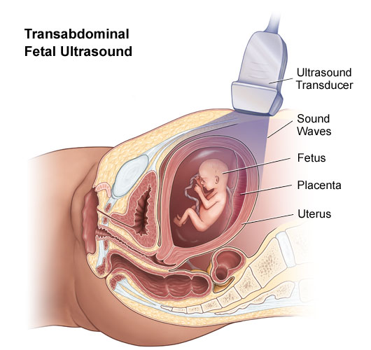
The amnion is a membrane that closely covers the embryo when first formed. It fills with the amniotic fluid which causes the amnion to expand and become the amniotic sac which serves to provide a protective environment for the developing embryo or fetus. The amnion is a membranous sac which surrounds and protects the embryo. The amniotic cavity is roofed in by a single stratum of flattened, ectodermal cells, the amniotic ectoderm, and its floor consists of the ectoderm of the embryonic disc. A thin layer of mesoderm, continuous with that of the somatopleure, is located just outside the amniotic ectoderm, and is connected to the mesodermal lining of the chorion by the body-stalk. When first formed, the amnion is in direct contact with the body of the embryo, but about the fourth or fifth week, amnionic fluid begins to accumulate within it. As the volume of the fluid increases, the amnion expands and ultimately adheres to the inner surface of the chorion.

Image result for amnion

For More Views: http://www.sciaeon.org/womens-health-and-complications/home        

Article page: http://www.sciaeon.org/womens-health-and-complications/articles-in-press

Contribute your manuscript: http://www.sciaeon.org/submit-paper

For more queries: whc@sciaeonopenaccess.com

Menarche

Menarche is the first menstrual cycle, or first menstrual bleeding, in female humans. From both social and medical perspectives, it is often considered the central event of female puberty, as it signals the possibility of fertility. Girls experience menarche at different ages. The timing of menarche is influenced by female biology, as well as genetic and environmental factors, especially nutritional factors. The time in a girl’s life when menstruation first begins. During the menarche period, menstruation may be irregular and unpredictable. Also known as female puberty.

For More Views: http://www.sciaeon.org/womens-health-and-complications/home        

Article page: http://www.sciaeon.org/womens-health-and-complications/articles-in-press

Contribute your manuscript: http://www.sciaeon.org/submit-paper

For more queries: whc@sciaeonopenaccess.com

Mammary Glands

Glandular organ located on the chest. The mammary gland is made up of connective tissue, fat, and tissue that contains the glands that can make milk. Also called breast. The mammary gland is a gland located in the breasts of females that is responsible for lactation, or the production of milk. Both males and females have glandular tissue within the breasts; however, in females the glandular tissue begins to develop after puberty in response to estrogen release. Mammary glands only produce milk after childbirth. During pregnancy, the hormones progesterone and prolactin are released. The progesterone interferes with prolactin, preventing the mammary glands from lactating. During this time, small amounts of a pre-milk substance called colostrum are produced. This liquid is rich in antibodies and nutrients to sustain an infant during the first few days of life. After childbirth, progesterone levels decrease and the levels of prolactin remain raised. This signals the mammary glands to begin lactating. Each time a baby is breastfed the milk is emptied from the breast. Immediately afterward, the mammary glands are signaled to continue producing milk.

Image result for mammary glands

For More Views: http://www.sciaeon.org/womens-health-and-complications/home        

Article page: http://www.sciaeon.org/womens-health-and-complications/articles-in-press

Contribute your manuscript: http://www.sciaeon.org/submit-paper

For more queries: whc@sciaeonopenaccess.com

Perineum

The perineum is generally defined as the surface region in both males and females between the pubic symphysis and the coccyx. The perineum is below the pelvic diaphragm and between the legs. It is a diamond-shaped area that includes the anus and, in females, the vagina. You can develop an abscess directly on the perineum or on a nearby area, such as the vulva or scrotum. An anal abscess can also cause pain in the perineum. These are usually the result of an infection of your internal anal glands. The perineum is the space between the anus and scrotum in the male and between the anus and the vulva in the female. The perineum is the region of the body between the pubic symphysis (pubic arch) and the coccyx (tail bone), including the perineal body and surrounding structures. There is some variability in how the boundaries are defined. The perianal area (peri- and anal) is a subset of the perineum. The perineum is an erogenous zone for both males and females. Perineal tears and episiotomy often occur in childbirth with first-time deliveries, but the risk of these injuries can be reduced by preparing the perineum, often through massage.

Image result for perineum

For More Views: http://www.sciaeon.org/womens-health-and-complications/home        

Article page: http://www.sciaeon.org/womens-health-and-complications/articles-in-press

Contribute your manuscript: http://www.sciaeon.org/submit-paper

For more queries: whc@sciaeonopenaccess.com

Fimbriae

The fimbriae of the uterine tube, also known as fimbriae tubae, are small, fingerlike projections at the end of the fallopian tubes, through which eggs move from the ovaries to the uterus. The fimbriae are connected to the ovary. Small epithelial cells — those that line cavities throughout the body — with small, slender cilia (microscopic, hair-like structures) pulsate inside the fallopian tubes to guide the ovum, or egg, from the ovary to the uterus. As there is no direct connection between the ovaries and fallopian tubes (also known as uterine tubes or oviducts), the egg is transported to the uterus in a peritoneal fluid produced by the fimbriae on the edge of the tube’s opening. Because the ovum cannot move by itself, the sweeping movement of the cilia of the fimbriae dictates its movement. It generally takes about 3 to 5 days for an egg to leave the ovary and land in the uterus.

Image result for fimbriae

For More Views: http://www.sciaeon.org/womens-health-and-complications/home        

Article page: http://www.sciaeon.org/womens-health-and-complications/articles-in-press

Contribute your manuscript: http://www.sciaeon.org/submit-paper

For more queries: whc@sciaeonopenaccess.com

Episiotomy

An episiotomy is an incision through the vaginal wall and the perineum to enlarge the vaginal opening and facilitate childbirth. During a vaginal birth, the health care provider will assist the fetus’ head and chin out of the vagina when it becomes visible. Once the head is out of the vagina, the shoulders are eased out, followed by the rest of the body. In some cases, the vaginal opening does not stretch enough to accommodate the fetus. In this case, an episiotomy may be done to help enlarge the opening and deliver the fetus. The episiotomy is usually done when the fetal head has stretched the vaginal opening to several centimeters during a contraction. Although episiotomy was a very common procedure in the past, more recent studies have found that routine or preventive use of episiotomy does not benefit the health of mother or baby.

Image result for episiotomy

For More Views: http://www.sciaeon.org/womens-health-and-complications/home        

Article page: http://www.sciaeon.org/womens-health-and-complications/articles-in-press

Contribute your manuscript: http://www.sciaeon.org/submit-paper

For more queries: whc@sciaeonopenaccess.com

Fetal ultrasound

Fetal ultrasound is a test used during pregnancy that creates an image of the fetus in the mother’s uterus, or womb. During a fetal ultrasound, various parts of the baby, such as the heart, head, and spine, are identified and measured. The testing may be performed either through the mother’s abdomen (transabdominal) or vaginal canal (transvaginal). Fetal ultrasound provides a safe way to evaluate the health of an unborn baby. Ultrasound uses an electronic device called a transducer to send and receive sound waves. When the transducer is moved over the abdomen, the ultrasonic sound waves then move through the skin, muscle, bone, and fluids at different speeds. The sound waves bounce off the fetus like an echo, returning to the transducer. The transducer picks up the reflected waves and converts them into an electronic picture. A clear gel is placed between the transducer and the skin to allow for the best sound conduction and smooth movement of the transducer.

Image result for fetal ultrasound

For More Views: http://www.sciaeon.org/womens-health-and-complications/home        

Article page: http://www.sciaeon.org/womens-health-and-complications/articles-in-press

Contribute your manuscript: http://www.sciaeon.org/submit-paper

For more queries: whc@sciaeonopenaccess.com

Loop Electrosurgical Excision Procedure

LEEP stands for Loop Electrosurgical Excision Procedure. It’s a treatment that prevents cervical cancer. A small electrical wire loop is used to remove abnormal cells from your cervix. LEEP surgery may be performed after abnormal cells are found during a Pap test, colposcopy, or biopsy. This procedure allows your physician to remove abnormal cell growth on the surface of the cervix and, if necessary, test it for cancer. LEEP is prescribed after the abnormal changes are confirmed by Pap tests and colposcopy. LEEP is an abbreviation for loop electrosurgical excision procedure. It is a way to test and treat abnormal cell growth on the surface tissue of the cervix. LEEP may be recommended after abnormal changes in the cervix are confirmed by Pap tests and colposcopy biopsies. Colposcopy is a non-invasive procedure in which a device similar to a microscope is used to view the cervix. LEEP allows your physician to remove the abnormal tissue and test it for cancer.

Image result for loop electrosurgical excision procedure

For More Views: http://www.sciaeon.org/womens-health-and-complications/home        

Article page: http://www.sciaeon.org/womens-health-and-complications/articles-in-press

Contribute your manuscript: http://www.sciaeon.org/submit-paper

For more queries: whc@sciaeonopenaccess.com

Postpartum Care

Pregnancy changes your body in more ways than you might expect, and it doesn’t stop when the baby is born. Here’s what to expect physically and emotionally after a vaginal delivery.
Vaginal soreness: If you had an episiotomy or vaginal tear during delivery, the wound might hurt for a few weeks. Extensive tears might take longer to heal. To ease discomfort while you’re recovering: Sit on a pillow or padded ring, Cool the wound with an ice pack, or place a chilled witch hazel pad between a sanitary napkin and the wound, Use a squeeze bottle to pour warm water on your perineum as you’re passing urine, Sit in a warm bath just deep enough to cover your buttocks and hips for five minutes. Use cold water if you find it more soothing, Take an over-the-counter pain reliever. Ask your health care provider about a numbing spray or cream, if needed, Talk to your health care provider about using a stool softener or laxative to prevent constipation.

For More Views: http://www.sciaeon.org/womens-health-and-complications/home        

Article page: http://www.sciaeon.org/womens-health-and-complications/articles-in-press

Contribute your manuscript: http://www.sciaeon.org/submit-paper

For more queries: whc@sciaeonopenaccess.com

Sonohysterography

Sonohysterography is also used to investigate uterine abnormalities in women who experience infertility or multiple miscarriages. Doppler ultrasound images can help the physician to see and evaluate: blockages to blood flow. blood flow in polyps, tumors and congenital malformation. Sonohysterography is a special kind of ultrasound exam. Fluid is put into the uterus through the cervix using a thin plastic tube. Sound waves are then used to create images of the lining of the uterus. The fluid helps show more detail than when ultrasound is used alone. Sonohysterography can find the underlying cause of many problems, including abnormal uterine bleeding, infertility, and repeated miscarriage. It is able to detect the following: Abnormal growths inside the uterus, such as fibroids or polyps, and information about their size and depth, Scar tissue inside the uterus, Abnormal uterine shape, Problems with the lining of the uterus, Whether the fallopian tubes are open or blocked.

For More Views: http://www.sciaeon.org/womens-health-and-complications/home        

Article page: http://www.sciaeon.org/womens-health-and-complications/articles-in-press

Contribute your manuscript: http://www.sciaeon.org/submit-paper

For more queries: whc@sciaeonopenaccess.com